
Diagnosing a Baker's Cyst
When it comes to diagnosing a Baker's Cyst, there are several different methods your doctor may use. This includes a physical examination, a review of your medical history, an x-ray, an MRI, an ultrasound and transillumination.

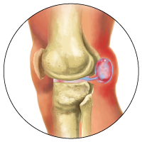
Physical Examiation - First of all, your doctor will examine your knee and leg to see if there are signs of swelling in the area of the Popliteal Bursa (the back of your knee), comparing the affected knee to the healthy one. Your doctor will probably feel around for inflammation and test the mobility of the affected knee. Your doctor will want to rule out other conditions which can present the same as a Baker's Cyst, such as a blood clot, tumor or aneurysm.

Medical History - Your doctor will want to review your medical history to see if you have any conditions that are related to a Baker's Cyst, such as arthritis or gout. You should also inform your doctor of any recent injuries to the knee or leg, as this could be what has caused your Baker's Cyst to develop.

X-Ray - Your doctor may want to conduct an x-ray exam of your knee if arthritis is suspected. A Baker's Cyst, however, would not show up on an x-ray.
MRI - An MRI (magnetic resonance imaging) exam would help your doctor check for any issues arising from complications with a suspected Baker's Cyst, such as a quickly growing cyst or symptoms of fever. An MRI may also be done to conclusively diagnose a Baker's Cyst, as it would show up clearly and help your doctor determine if there is cartilage damage.

Transillumination - This is another non-invasive test that involves shining a light through your Baker's Cyst to see if there is fluid inside.
Ultrasound - An ultrasound may need to be done to diagnose the Baker's Cyst or to check for any damage to the knee that could be causing your Baker's Cyst.